 |
| SimpsonWorld [Internet] [Cited 2021 June 06] Available from; https://giphy.com/gifs/season-8-the-simpsons-8x2-3orifb9uia6g30MbE4 |
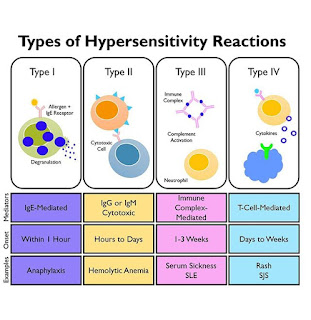
The traditional classification for hypersensitivity reactions is that of Gell and Coombs and is currently the most commonly known classification system. [1]
It divides the hypersensitivity reactions into the following 4 types:
⭐Type I hypersensitivity (Allergy Immediate Anaphylactic)
Also known as an immediate reaction and involves immunoglobulin E (IgE) mediated release of antibodies against the soluble antigen. This results in mast cell degranulation and release of histamine and other inflammatory mediators.
⭐Type II hypersensitivity (Cytotoxic, Antibody-dependent)
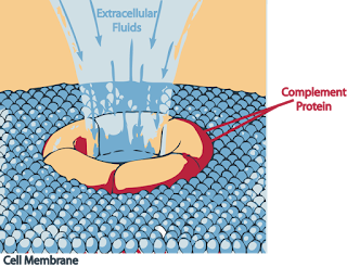
Also known as cytotoxic reactions and engages IgG and IgM antibodies, leading to the complement system activation and cell damage or lysis.
⭐Type III hypersensitivity (Immune complex)
Also known as immune complex reactions and involves IgG, IgM, and sometimes IgA antibodies. The build-up of these immune complexes results in complement system activation, which leads to polymorphonuclear leukocytes (PMNs) chemotaxis and eventually causing tissue damage.
⭐Type IV hypersensitivity (Immune Delayed, cell-mediated immune memory response, Antibody-independent)
Also known as delayed-type and involves of T-cell-mediated reactions. T-cells or macrophages are activated as a result of cytokine release, leading to tissue damage. (2)
 |
| Diaz G. GrepMed [Internet] 2019 [Cited 2021 June 06] Available from; https://www.grepmed.com/images/6381/hypersensitivity-diagnosis-classification-reactions-types |
Type I Hypersensitivity
 |
| Grammartop[Internet] 2020 Nov. 17 [Cited 2021 June 06] Available from; https://grammartop.com/allergy-synonyms/ |
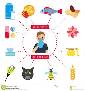
The Type I hypersensitivity is due to aberrant production and activity of IgE against normally nonpathogenic antigens (commonly called allergens) This allergic hypersensitivity is also called immediate hypersensitivity because of the speed of reaction development. (5)
There are two categories in Type I hypersensitivity diseases,(2)
1. Atopic diseases ( an exaggerated IgE mediated immune responses )
- Allergic: asthma,
- Rhinitis,
- Conjunctivitis,
- Dermatitis

Atopic Dermatitis Silverberg J. Comorbidities and the impact of atopic dermatitis [Internet] 2019 Aug. 01 [Cited 2021 June 07] Available from; DOI:https://doi.org/10.1016/j.anai.2019.04.020
2. Allergic diseases (immune responses to foreign allergens)
- Anaphylaxis,
- Urticaria,
- Angioedema,
- Food, and drug allergies (2)
 |
| Allergic Reactions AMBOSS[Internet][Cited 2021 June 07] Available from; https://www.amboss.com/us/knowledge/Hypersensitivity_reactions |
The allergens that result in a type I hypersensitivity may be harmless (i.e., pollen, mites, or foods, drugs, etc.) or more hazardous such as insect venoms(2).
The risk factors of Type I Hypersensitivity
- Geographical distribution,
- Environmental risks such as pollution or socioeconomic status,
- Genetic predisposition, or the “hygiene hypothesis”(2).
Hygiene hypothesis
The hygiene hypothesis states that allergy and increased longevity are both consequences of reducing infectious stressors during early childhood. It suggests that our modern society practices of good hygiene and the lack of early exposure to many microbes or antigens may result in failures of the immune system functionality.As such, the hypothesis suggests that early exposure to a diverse range of microorganisms and antigens may actually lead to overall decreased rates of allergies, asthma, and other immune disorders(3).
Hygiene/"Old friend" Hypothesis
Etiology of Type I Hypersensitivity
The responses of Type I hypersensitivity occurs due to antigens And these responses to Antigen occurs in two stages
1. Sonication
2. Effect stage
Diagnosing of Type I Hypersensitivity
In-Vitro testing- Serum tryptase, which is released by mast cells in an hour post-activation, elevated levels can indicate an increased risk of anaphylaxis.
- Eosinophil count can be increased in atopic disorders.
- Allergen-specific IgE assays are preferable to in-vivo skin testing, as they can help identify food allergens or other antigens that are causing a reaction.
- Histamine levels may be collected; however, the assays are not reliable, and the levels are short-lived.
- Flow cytometric basophil activation test (BAT) can help diagnose immediate hypersensitivity to certain medications
- Skin Test
- Drug provocation test (DPT),
- Spirometry or pulmonary function tests can be used to assess asthma.
- Inhalation challenges with specific allergen and histamine, mannitol, and methacholine are used to assess airway hypersensitivity.
- Eosinophil count from nasal smear or induced sputum can be collected as well to confirm hypersensitivity. (2)
Management and Treatment of Type I Hypersensitivity
1)Anaphylaxis
- Epinephrine
- Bronchodilators
- Antihistamines: i.e., diphenhydramine, famotidine or ranitidine
- Glucocorticoids
2)Urticaria/Angioedema
- Antihistamine
- Glucocorticoids
3)Allergic Asthma
- Inhaled beta-agonists with or without inhaled corticosteroids
4)Allergic Rhinitis
- Topical nasal or optical decongestants
5)Allergic Conjunctivitis/Allergic Dermatitis/Eczema/Wasp or bee venom/Drug Allergy/Food Allergy
- Oral or topical H1 antihistamine and oral or inhaled glucocorticoids may be used for symptomatic control. (2)
 |
| Medineplus [Internet][Cited 2021 June 09] Available from; https://medlineplus.gov/ency/article/000812.htm |
Type II Hypersensitivity
 |
| Creative_Med_Dose [Internet][Cited 2021 June 09] Available from; https://creativemeddoses.com/topics-list/type-ii-hypersensitivity-antibody-mediated/ |
Antigen of Type II Hypersensitivity are present or is a part of the cell membrane. It may be:
Exogenous like Microbes, parasites, drugs or Intrinsic like Autoimmune diseases, and these are self-Antigens
- Antibody binding to cell surface receptors and altering its activity
- Activation of the complement pathway.
- Antibody dependent cellular cytotoxicity (6)
 |
| Complement Death Swilliams, Complement death [Internet] WikiDoc,2008 May 21 [Cited 2021 June 10] Available from; https://www.wikidoc.org/index.php/File:Complement_death.PNG |
Etiology of Type II Hypersensitivity
Causes and Disorders of Type II hypersensitivity
- Penicillin,
- Cephalosporins,
- Hydrochlorothiazide,
- Methyldopa
- Idiopathic thrombocytopenic purpura (platelet destruction)
- Pemphigoid reactions
- Myasthenia gravis (muscle weakness)
- Graves' disease (hyperthyroidism)
- There is an extreme weakness in the muscles.
- There are autoantibodies against the acetylcholine receptor present on the surface of the muscle membrane.
- The acetylcholine receptors are present at the motor endplate, where the neuron contacts the muscles.
- Where Ab reacts with acetylcholine receptors.
- There is the transient weakness of the newborn babies because of the mother’s passive transfer (myasthenia gravis patient) IgG antibodies(8).
 |
| Myasthenia gravis Creative_Med_Dose [Internet][Cited 2021 June 09] Available from; https://creativemeddoses.com/topics-list/type-ii-hypersensitivity-antibody-mediated/ |
- Blood transfusion reactions: This may take place because of mismatched blood transfusion.
- Hemolytic diseases of the newborn (HDN), e.g., Rh-incompatibility and ABO-Incompatibility.
- Autoimmune hemolytic anemia (AHA). This takes place due to the loss of immunologic tolerance.
- It can be divided into three main types:
- Warm-reactive autoantibody, these autoantibodies react with antigen at 37 °C.
- Cold reactive autoantibodies react with antigen below 37 °C and mostly at 4 °C.
- These antibodies are provoked by the drugs causing an allergic reaction.
- The possibilities of RBC Ag and Ab reactions are:
- Agglutination.
- Complement dependent lysis.
- Phagocytosis (8)
Testing of Type II hypersensitivity
The testing includes the following:
1). Complete blood cell count surveying;
- Hemoglobin (autoimmune hemolytic anemia),
- Red blood cell count,
- Neutrophils,
- Lymphocytes,
- and platelets for cytopenia
2). An assessment of immunoglobulins with a quantitative assay for IgG, IgM, and IgE depending on the type of hypersensitivity reaction.
3). Animals, plants, food, and environmental pollutants can be used in skin prick tests
4). A radioallergosorbent test (RAST for IgE)
5). The detection of autoantibodies can be assessed in systemic autoimmune diseases like SLE and rheumatoid arthritis (RA).
6). Autoantibodies can also be done against specific organs,
- Example- autoantibody against islet cells in diabetes mellitus, thyroid-stimulating hormone (TSH) receptor antibody to differentiate it from other causes of thyrotoxicosis, and acetylcholine receptor antibody test (highly specific for myasthenia gravis). (6)
 |
| Laboratory Tests Domocmat C. Hypersensitivity reactions [Internet] 2013 Feb. 03 [Cited 2021 June 09] Available from; https://www.slideshare.net/kharr/hypersensitivity-reactions-cld |
Treatment and Management of Type II hypersensitivity
- Antibiotic therapy should be started to eradicate the carriage of group A streptococcus-Penicillin V and benzylpenicillin are primarily used for this purpose.
- Oral cephalosporins are used as an alternative therapy for penicillin-allergic patients.
- Symptomatic treatment of acute manifestations like;
- Arthritis (with NSAIDs),
- Carditis (with aspirin or oral prednisone),
- and chorea (with diazepam)
- Anti-thyroid drugs, propylthiouracil and methimazole,
- Radioiodine ablation and surgical excision should also be considered.
- Glucocorticoids are used for ophthalmopathy.
- Acetylcholinesterase inhibitors like pyridostigmine are started.
- Immunosuppressive therapy (like corticosteroids) is used for the patients refractory to pyridostigmine therapy.
- Thymectomy is considered in patients refractory to drug therapy.
- Plasma exchange and intravenous immune globulin should be given in myasthenic crisis. (6)


Comments
Post a Comment